Dental X-Ray and Orthopantqmogram (OPG) in Pune
Dental X-Ray in Pune
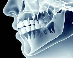
Dental xray (Radiograph) is the image of one or two teeth that your dentist uses to exaluate the condition of your teeth. Dental xrays are taken using a film, a scanner and Xray cone. A film is kept against the concerned tooth and xray beam is shot toward tthe film. This captures the image of the teeth on the film, which is later developed using a scanner or other conventional techniques like using a chemical developer. Dental xray helps to diagnose the underlying disease associated with the teeth, bone and surrounding critical structures. Usually a radiograph is taken to asscess the periodontium of the tooth like the periodontal ligament and the Bone. Any chnages in the periodonrium due to infection is directly reflected in the xray of that particular tooth.
A periodontally coompromised tooth will show widened periodontal ligament, resorbed bone, rareified osseous structure. Dental caries are clearly evident in a dental xray. The depth of dental caries can be assessed using a radiograph, which will fiurther help in treatment planning. Also dental xray helps to have a more defined look at the impacted terth.
Talk with Our Expert Dentist for Dental Xray at Pathak Dental Clinic, Pimpri-Chinchwad, Pune
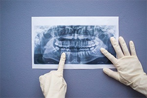
Orthopantamogram (OPG)
OPG is used to create image of the entire mouth. All the structures like teeth, alvelolar bone, Temporomandibular Joint, maxillary sinus, zygomatic bone,, impacted teeth(usually the upper and llower impacted third molars ), and any anomalies related to these vital structures are seen. OPG is of a great use in planning orthodontic treatments. How is an OPG taken? The patient is made to stand near an opg machine and the chin of the patient is made to rest upon a stand. Then tthe ptient is asked to bite in full occlusion. The arm of the OPG machine revolves are the patients head and takes several images which are then merged in the software to produce a final picture. OPG has a disadvantage of forming ghost images due to overlap of structures like tongue.
Talk with Our Expert Dentist for Orthopantamogram (OPG) at Pathak Dental Clinic, Pimpri-Chinchwad, Pune
How safe is Dental X-Ray and OPG?
Your body can be exposed to 3.1 millisieverts of natural radiation per year. A dental xray exposure is less than 0.005 millisiverts which is less than 16% of ur daily natural exposure. Also we habe a AERB approval liscense to operate a dental xray unit. Patients are asked to wear a thyroid collar or a lead apron to avoid unnecessery radiation exposure.
Dental X-ray treatment cost is Rs. 200.
Frequently Asked Questions
Q.Are Dental X-Rays Dangerous?
Many people wonder if dental X-rays are dangerous or could potentially cause harm. The truth is, that when administered by a trained and licensed professional, they are incredibly safe.
Q.What are the benefits of Dental X-ray and OPG?
Some of the benefits of Dental X-ray and OPG are:
- Low patient radiation dose.
- Better coverage of teeth and facial bones.
- Convenience in examining a patient's oral health.
- Beneficial for patients who have restricted jaw movements.
- Simple and effective process to produce oral images.
- Provides visual aid while educating patients and presenting cases.
Q.Why is an OPG or Dental X-ray done?
Dental X-rays (radiographs) are images of your teeth that your dentist uses to evaluate the overall oral health or identify specific problems, like cavities, tooth decay, and impacted teeth. OPG is used by dentists to view all their patient’s teeth and including their number, position, and growth, including those that have not yet erupted.
Q.Can OPG detect cavity?
An orthopantomogram (OPG) is a common radiograph used to identify the hard tissues of the oral cavity and surrounding skeletal structures.
About Author

Dr.Manish Pathak
BDS, MDS, Owner of Pathak Dental Clinic
Dr.Manish Pathak pursed his BDS and MDS From Maharashtra university of health science. He has various national and international publications on his name and in the last 8 years. He received many National and International awards in the field of dentistry. Currently, Dr.Manish Pathak Runs his private dental clinic in Pune and consultant over 100 clinics across Pune.